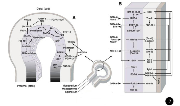
Figure 4 .
Ways and means by which prominent signaling molecules and transcription factors control lung development. The middle (small) diagram shows the different parts (germ layers) of a lung bud (proximal and distal). Figure 7A: Location of signalling molecules and the pathways by which they control growth, budding, and differentiation of the airway epithelium and surrounding mesenchyme. The shading shows the proximal-distal extent of the lung bud epithelium. Figure 7B: Signaling programs in the distal lung bud bring about mesenchymal- and epithelial cell proliferation as well as airway branching. Details to these processes can be obtained from the text and particularly from the following comprehensive reviews: Metzger and Krasnow [25], Perl and Whitsett [11], Roth-Kleiner and Post [116], Cardoso and Lü [40], Lu and Werb [53], De Langhe and Reynolds [117], Affolter et al. [77], Warburton et al. [21], [15,16,18,29,98]. Modified after Ornitz and Yin [18].

