Figure 8 .
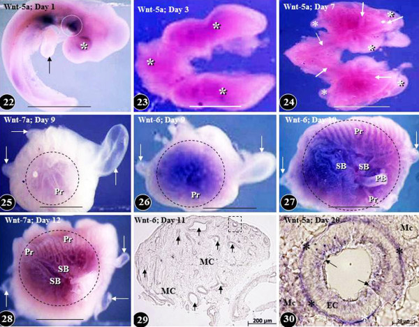
Expression of Wnt signaling molecules in developing chicken embryo (Figure 22) and in the lung at different times of embryonic life (Figures 23–30). Wnt-5a is expressed on the ventral aspect of the body trunk at day two of life (Figures 22). Arrow, developing fore limb bud; asterisk, optic placode; dashed circle, prospective site of the development of the lung and the heart. Scale bar, 1 mm. Figures 23: Diffuse expression of Wnt-5a (asterisks) in the developing lung on day three of life. Scale bar, 1 mm. Figure 24: Expression of Wnt-5a in developing lung on day five of life. Wnt-5a is predominantly localized in the developing airways (arrows) and relatively little of it in the mesenchyme and the air sacs (asterisks). Scale bar, 1 mm. Figures 25–28: Expression of Wnts in developing lungs at different embryonic days of life (areas marked by dashed circles). Figures 25, 26, and 28 show lateral aspects of the lung while Figure 27 is that of a medial one. The Wnts are poorly expressed in the developing air sacs (arrows). PB, primary bronchus; Pr, parabronchi; SB, secondary bronchi. Scale bars, 1 mm. Figure 29: Transverse section of a developing lung at the eleventh day of life showing Wnt-6 expression in the epithelium lining the airways (arrows) and the mesenchyme (MC). An airway like the one enclosed in the dashed square is shown enlarged in Figure 30. Figure 30: Cross-section of a parabronchus showing the expression of Wnt-5a at the 20th day of development. Wnt-5a is highly expressed in the epithelial cells (arrows), the mesenchymal cells (MC), and in the basement membrane of the epithelial cells (stars). Figures 22–28 are from Maina [132]; Figure 30 is from Maina [134]; Figure 29 is from unpublished work of RG Macharia and JN Maina.

