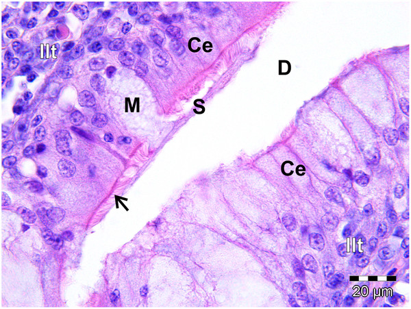
Figure 10.

Ciliated columnar lymphoepithelium (Ce) lining a gland in the follicular pharyngeal tonsil of S. camelus. The mucus-secreting gland has been invaded by lymphocytes of the inter-nodular lymphoid tissue (Ilt). The cilia (arrow) spread the secreted mucus (S) from the mucus cell (M) into the gland duct (D).
