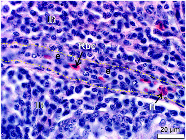
Figure 14.

A venule traversing the inter-nodular lymphoid tissue (Ilt) in the follicular pharyngeal tonsil of S. camelus. The wall of the venule is roughly outlined (yellow dotted lines) for clarity. The endothelial nuclei (e) are pale and oval to round. Note the high number of lymphocytes (*) within the lumen. Red blood cell (Rbc) and heterophil (H).
