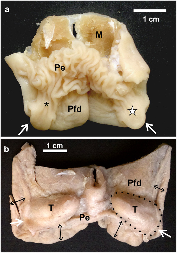
Figure 2.

Dorsal view of the pharyngeal folds and associated follicular tonsils. a: D. novaehollandiae. The pharyngeal tonsil (white star) is situated entirely within the recess formed between the dorsal, free surface of the pharyngeal folds (Pfd) and the proximal esophagus (Pe) which is continuous with the base of the caudo-lateral extension (*). The opening of the tonsil to the oropharynx is indicated (white arrows). Muscle tissue (M). b: S. camelus. The opening of the pharyngeal tonsils to the oropharynx is indicated (white arrows). The proximal esophagus has been removed to expose the retropharyngeally positioned pharyngeal tonsils (T) (black dotted outline) as well as the dorsal aspect of the short free part (black double-headed arrows) of the pharyngeal folds (Pfd).
