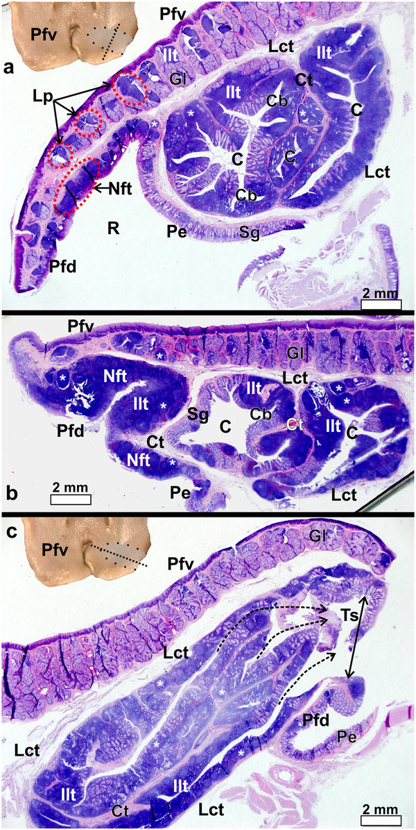
Figure 6.

Pharyngeal fold and pharyngeal tonsil of S. camelus. a-b: Transverse sections as indicated by the dotted line in the inset. Note the numerous simple, branched, tubular mucus-secreting glands (Gl) and associated lymphoid tissue surrounded by connective tissue (Ct) present on the ventral surface of the pharyngeal fold (Pfv). Lymphnoduli pharyngeales (Lp) are associated with the ventral surface of the folds. The dorsal surface of the pharyngeal fold (Pfd) displays substantial amounts of both lymph nodules (*) and inter-nodular (Ilt) lymphoid tissue which in places forms a non-follicular pharyngeal tonsil (Nft). The follicular pharyngeal tonsil is formed by a concentration of lymph nodules and inter-nodular lymphoid tissue positioned between the ventral surface of the pharyngeal fold and the proximal esophagus (Pe) and is encapsulated by loose connective tissue (Lct). The proximal esophagus, the dorsal surface of the pharyngeal fold and the tonsil all contain simple, tubular mucus-secreting glands (Sg) which are invaded to varying degrees by lymphoid tissue. Recess (R) between the dorsal surface of the pharyngeal fold and proximal esophagus, tonsillar crypts (C) and branches of the crypts (Cb). c: Longitudinal section as indicated by the dotted line on the inset. Note the numerous crypts which open via fossules (dotted arrows) into the shallow tonsillar sinus (Ts) (double-headed arrow) before opening into the oropharynx. The pharyngeal tonsil is separated from the pharyngeal fold by a loose connective tissue capsule (Lct). Simple, branched, tubular mucus-secreting glands (Gl), ventral (Pfv) and dorsal (Pfd) surfaces of the pharyngeal fold, lymph nodules (*) and inter-nodular (Ilt) lymphoid tissue, connective tissue (Ct) and proximal esophagus (Pe).
