Figure 9.
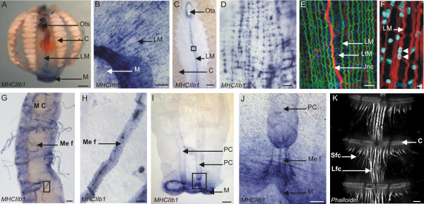
PpiMHCIIb1 expression outside the tentacle root. (A) General view of the PpiMHCIIb1 expression pattern. (B) Detail of the expression of PpiMHCIIb1 in longitudinal parietal muscle fibres around the mouth. (C) Expression of PpiMHCIIb1 in longitudinal parietal muscle fibres located in the tentacular plane, between the tentacle sheath opening and the mouth. (D) Higher magnification of the region boxed in (C). (E) Phalloidin (in green), YL1/2 (in red) and DAPI (in blue) staining of the same region as shown in panel (D) but from another specimen. (F) Phalloidin (in red) and DAPI (in blue) staining showing the multinucleation of parietal muscle fibre. (G) Expression of PpiMHCIIb1 in mesogleal muscle fibres (Me F) connected to a meridional canal (M C). (H) Higher magnification of one of these PpiMHCIIb1 expressing mesogleal muscle fibres boxed in (G). (I) Expression of PpiMHCIIb1 gene in the mouth rim (M) and in mesogleal muscle fibres connecting the two paragastric canals (PC) to the mouth. (J) Higher magnification of the region boxed in (I) to show the stained mesogleal muscle fibres (Me f) connecting a paragastric canal (PC) to the mouth. (K) Portion of a comb row (with 3 successive combs) showing the inter-comb fibres, strongly stained with phalloidin. There are two types of inter-comb fibres: short lateral fibres (Sfc) and long central fibres (Lfc). C: Comb; Jnc: Juxtatentacular nerve cord; Lfc: Long inter-comb fibre cells; LM: Longitudinal muscle fibres; LtM: Latitudinal muscle fibres. M: Mouth; MC: Meridional Canal; Me f: Mesogleal muscle fibre; Ots: Orifice of tentacle sheath; PC: Paragastric canal; Sfc: Short inter-comb fibre cells. Scale bars: A, C, I: 200 μm; B: 100 μm; D, K: 20 μm; E, H: 10 μm; F: 2 μm; G, J: 50 μm.

