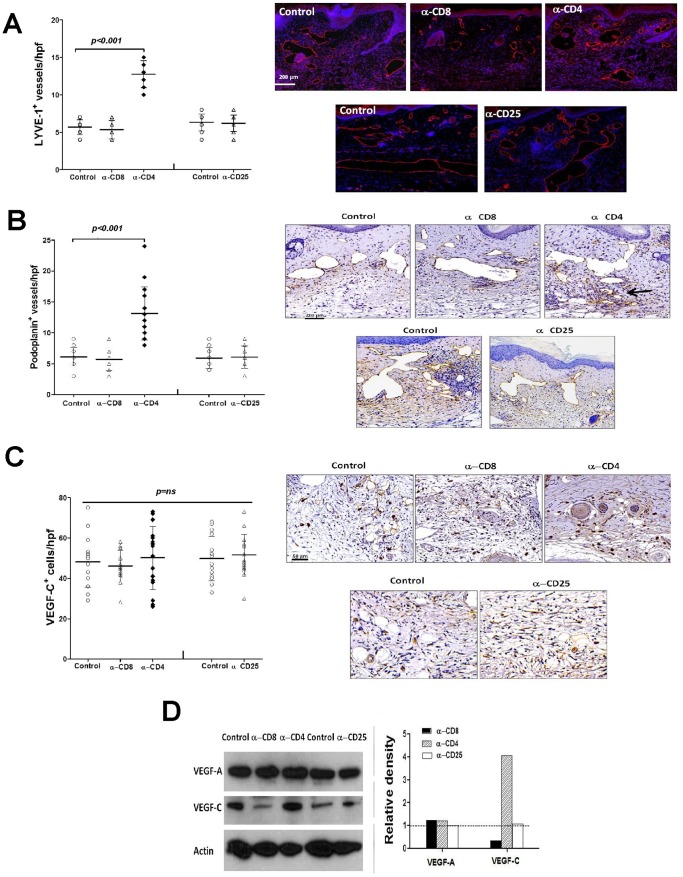
Figure 8. Loss of CD4+ but not CD8+ or CD25+ cells increases lymphangiogenesis. A.
LYVE-1+ vessel counts (left) and representative figures (right) of tail sections from control, CD8+, CD4+, or CD25+ cell depleted mice 6 weeks after surgery. B. Podoplanin+ vessel counts (left) and representative figures (right) of tail sections from control, CD8+, CD4+, or CD25+ cell depleted mice 6 weeks after surgery. C. VEGF-C+ cells/high powered field (hpf) counts (left) and representative figures of VEGF-C immunohistochemistry in tail tissues from control, CD8+, CD4+, or CD25+ cell depleted animals (right) 6 weeks after surgery. D. Representative (of triplicate experiments) western blot analysis of VEGF-A, and VEGF-C expression in protein lysates obtained from tail tissues from control, CD8+, CD4+, or CD25+ cell depleted animals 6 weeks after surgery. Quantification of band density relative to controls (arbitrarily set at 1 and represented by dotted line) is shown to the right.

