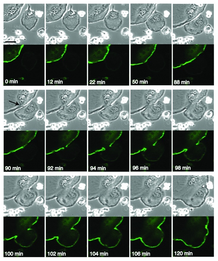
Figure 1. Cell fusion between EGFP-positive and EGFP-negative osteoclasts. RAW 264.7 cells were transfected with EGFP-actin and induced osteoclastogenesis by adding RANKL. The osteoclasts were processed for time lapse confocal microscopy as described.5 Images were retrieved every 2 min. Upper panel, DIC images. Lower panel, EGFP images. At 90 min, a stalk-like link appeared between two cells at the contact area (arrow). Bar, 50 µm.

An official website of the United States government
Here's how you know
Official websites use .gov
A
.gov website belongs to an official
government organization in the United States.
Secure .gov websites use HTTPS
A lock (
) or https:// means you've safely
connected to the .gov website. Share sensitive
information only on official, secure websites.
