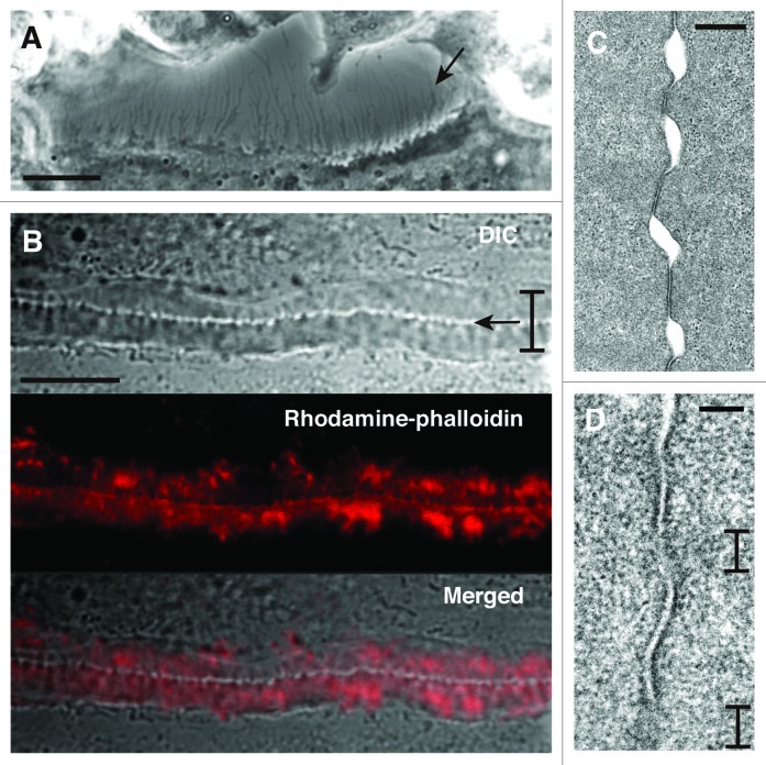
Figure 2. Fine structure at the site of cell contact between osteoclasts. (A) DIC image of osteoclast at day 4. The image shows the space between two osteoclasts. An osteoclast at lower position extends many thin processes at the cell surface (arrow). Bar, 10 µm. (B) The interface of two osteoclasts forms the zipper-like structure. Bracket indicates the short axis of the zipper-like structure. Arrow indicates the apparent physical links at the interface of two osteoclasts in the zipper-like structure. Upper, DIC image. Middle, rhodamine-phalloidin staining. Lower, merged image. Bar, 10 µm. (C) Transmission electron microscopy of the interface of two osteoclasts. Two osteoclasts attach to each other with gaps. Bar, 500 nm. (D) Apposed plasma membranes often contain fuzzy areas. Brackets indicate the fuzzy area. Bar, 100 nm.

An official website of the United States government
Here's how you know
Official websites use .gov
A
.gov website belongs to an official
government organization in the United States.
Secure .gov websites use HTTPS
A lock (
) or https:// means you've safely
connected to the .gov website. Share sensitive
information only on official, secure websites.
