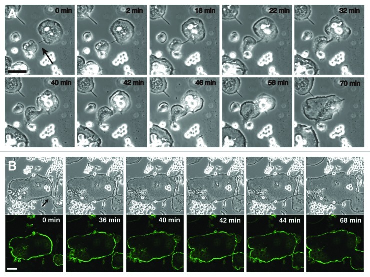
Figure 3. Filopodium-mediated fusion of osteoclats. (A) DIC imaging of the filopodium-mediated fusion between osteoclasts. RAW 264.7 cells were induced osteoclastogenesis by RANKL. Time-lapse recordings delineate the sequence of cell-cell recognition, an intermediate stage, fusion of the cell bodies, and the reshaping of the fused cell. See text for details. The arrow indicates the filopodium. (B) Dynamics of podosome rings during the filopodium-mediated fusion. RAW 264.7 cells were transfected with EGFP-actin and induced osteoclastogenesis by RANKL. At time 0 min, two osteoclasts were linked with a filopodium (arrow). Two podosome belts in the linked cells maintained their independence until the fusion of the cell bodies. Images were retrieved every 2 min. Upper panel, DIC images. Lower panel, EGFP images. Bars, 50 mm.

An official website of the United States government
Here's how you know
Official websites use .gov
A
.gov website belongs to an official
government organization in the United States.
Secure .gov websites use HTTPS
A lock (
) or https:// means you've safely
connected to the .gov website. Share sensitive
information only on official, secure websites.
