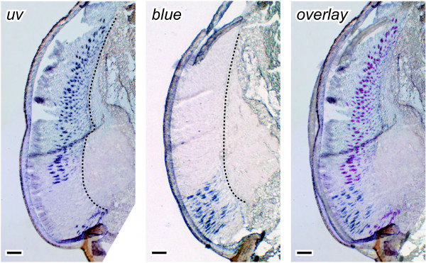
Figure 4.
Localization of uv and blue mRNA in the pigmented part of the compound eye. Consecutive dorso-ventral sections through the retina of an adult female were hybridized with antisense uv and blue riboprobes. The right panel shows an overlay of the two pictures to the left with the uv signals re-colored in purple. UV and blue opsin are transcribed in non-overlapping eye regions and at least partly at different levels of the ommatidia. The blue labeling is most intense distally, whereas uv is generally detected further proximally. Up corresponds to dorsal in all panels. Broken line = basement membrane, scale bars = 100 μm.

