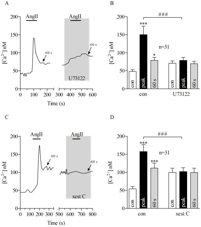
Figure 2. AngII-evoked Ca2+ response is mediated by PLC/IP3 pathway in porcine RPE cells.
Application of AngII for 80 seconds (bars) caused transient Ca2+response in pRPE cells. A: Bath application of 100 nM AngII (bars) together with 10 µM U73122 (gray shadow), a phospholipase C (PLC) blocker abolished AngII-evoked Ca2+signal in pRPE cells. On the left panels of Figures A and C, it is shown the effect of AngII application under control conditions. In the same cell, after washing out first AngII application (until [Ca2+]I returned back to basal levels) it was further perfused 100 nM of AngII in the presence of U73122 (right panel in A) or xest C (right panel in C). The wash out is not shown and the x-axis is interrupted accordingly between the panels. Note that application of U73122 alone led to a slight increase in intracellular free Ca2+. B: summary of data from experiments shown in A. C: Co-application of 100 nM AngII (bar) with the IP3 blocker xestospongin C (xest C) (gray shadow) at 10 µM reduced AngII-evoked Ca2+response in pRPE cells. D: summary of data from experiments shown in C. Bars in Fig. 2B and D represent means ± SEM for AngII-evoked Ca2+responses before (open bars) during the peak (black bars) and at 60 s after the maximum AngII-elicited calcium response (gray bars). *p<0.05, (***; ###) p<0.0001; repeated measures ANOVA. n = number of cells from 4 (B) or 5 (D) independent experiments.

