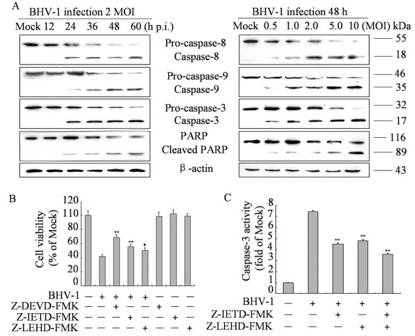
Figure 2.

Effects of BHV-1 infection on caspases and PARP cleavage in MDBK cells. (A) Western blot analysis of caspases activities and PARP cleavage in BHV-1-infected cells (2 MOI) for different times (left panel) or for different MOIs for 48 h p.i. (right panel). Data are representative of three separate experiments. β-actin was used as an internal loading control. (B) Effect of caspase inhibitors on cell viability. Cells were incubated with 20 μM of each caspase inhibitors for 2 h prior to infection with BHV-1 at 2 MOI for 48 h and cell viability was evaluated by MTT assay. Values are shown as mean ± SD, * p < 0.05, ** p < 0.01 versus BHV-1 infection alone without inhibitors. (C) The inhibitory efficacy of caspase-8 and −9 inhibitors on the activity of caspase-3. Cells were incubated with 20 μM of each caspase inhibitors for 2 h prior to infection with BHV-1 at 2 MOI for 48 h. the activity of caspase-3 was measured by colorimetric assay kit. The relative caspase-3 activity was calculated after the activity of caspase-3 in cells that treated with BHV-1 or inhibitors subtracted the background value. Values are mean ± SD. * p < 0.05, ** p < 0.01 versus BHV-1 infection alone without inhibitors.
