Figure 1.
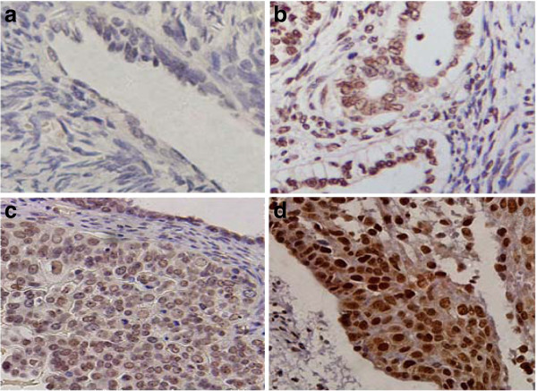
Expression of CIAPIN1 in EOC tissues and paired non-cancerous tissues by immunohistochemistry. ( a) Negative staining (−) of CIAPIN1 in paired non-cancerous tissues. ( b) Positive staining (+) of CIAPIN1 in the nucleus in well-differentiated tumor tissue. ( c) Moderate nuclear positive staining (++) of CIAPIN1 in moderately differentiated EOC tissues. ( d) Strong nuclear staining (+++) of CIAPIN1 in poorly differentiated EOC tissues. (Original magnification, 200×).

