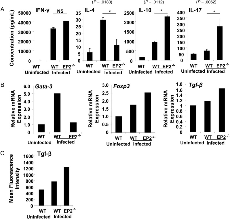
Figure 4.
EP2-deficient mice induce regulatory T cells upon infection with Mycobacterium tuberculosis. A, T helper 1 (IFN-γ), T helper 2 (IL-4 and IL-10), and T helper 17 (Th17) cytokine responses were assayed in the culture supernatants of splenocytes derived from uninfected and infected C57BL/6 (wild-type [WT]) and EP2−/− mice at 48 hours after activation with M. tuberculosis H37Rv protein lysate (complete soluble antigen [CSA], 20 μg/mL). Data shown here are representative of 3 independent experiments, with 3 mice per experiment. Abbreviations: IFN-γ, interferon γ; IL-4, interleukin 4; IL-10, interleukin 10; IL-17, interleukin 17; NS, not significant. B, EP2−/− mice show increased T regulatory cell–specific transcription factor (Foxp3) and cytokine (Tgf-β) expression. CD4+ T cells derived from uninfected and infected C57BL/6 (WT) and EP2−/− mice were analyzed for expression levels of Gata-3, Foxp3, and Tgf-β messenger RNA by real-time polymerase chain reaction. Data are representative of 2 experiments, with 3 mice per group. C, Infected EP2−/− mice show increased Tgf-β secretion in the culture supernatants of splenocytes rechallenged ex vivo with M. tuberculosis CSA. Data are representative of 2 experiments, with 3 mice per group.

