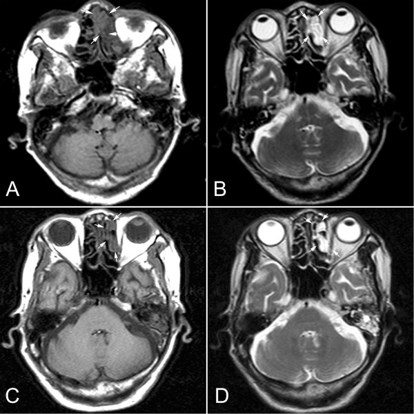
Figure 1.
Radiological findings of the intranasal mass (A) Axial CT scan (T1) revealed that an irregular mass presented in the left nasal cavity displacing the nasal septum (white arrow). (B) CT scan (T2) showed that the mass also extended into left ethmoid sinus, but did not erode the bony margins of the medial wall of the left orbit (white arrow). After 6-month period of follow-up, axial CT scan T1 (C) and T2 (D) showed that location and size of the mass did not change remarkably (white arrow).

