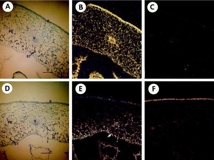
Figure 3.
Cross-sections of fruit approximately 10 mm in diameter were hybridized with the Frk1 probe in B and C, and with the Frk2 probe in E and F. Antisense probes were used in B and E, and sense (control) probes were used in C and F. Hybridization signals are visible as white dots in dark-field microscopy of the sections. The arrows in E indicate the concentrated signals of Frk2 transcripts. Bright-field microscopy of the sections hybridized with the antisense or sense probe are shown in A and D. The dyes used in staining these sections were toluidine blue and KI/I2. Bars = 200 μm; PE, pericarp; E, epidermis; V, vascular tissue; and ST, starch granule.

