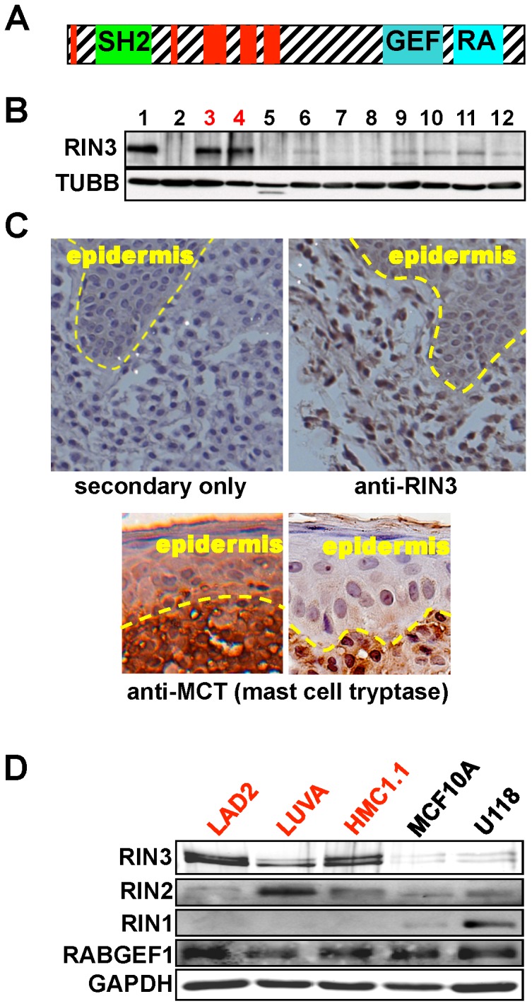
Figure 1. RIN3 is highly expressed and enriched in human mast cells.

(A) The domain structure of RIN3. Red = proline-rich motifs. (B) Immunoblot analysis of RIN3 expression in human cell lines. 1:NIH3T3 w/RIN3 (+ctr), 2:NIH3T3 (−ctr), 3:HMC1 (mast), 4:LAD2 (mast), 5:THP1 (macrophage), 6:SaOs2 (osteoclast), 7:RAMOS (B cell), 8:K562 (myoblast), 9:Jurkat (T cell), 10: MCF10A (epithelial), 11:IMR90 (fibroblast), 12:U118 (glioblastoma). RIN3 appears as a double or single band depending on exposure time of the immunoblot. TUBB = β-tubulin. (C) Human mastocytosis sections stained using immunohistochemistry for RIN3 or secondary only as a control. Sections stained with anti-mast cell tryptase are shown to identify the infiltrating mast cells. (D) Lysates from three human mast cell lines (LAD2, LUVA, HMC1.1), an epithelial cell line (MCF10A), and a neuronal cell line (U118) were run on an SDS-PAGE gel and immunoblotted for RIN family members (RIN1/2/3) as well as RABGEF1, another GEF for RAB5.
