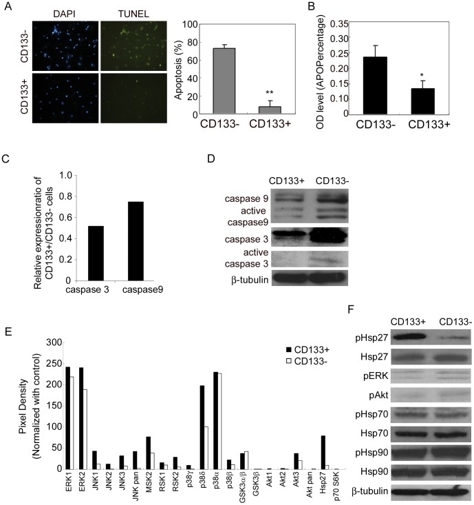
Figure 3. CD133+ cells resist hypoxia and serum depletion-induced apoptosis via activation of Hsp27.
(A) TUNEL staining for apoptosis was performed after MACS separation of CD133− and CD133+ cells under hypoxia and serum depletion for 4 days. (B) CD133− and CD133+ cells under hypoxia and serum depletion for 3 days were harvested by MACS separation, reseeded under hypoxia and serum depletion, and APOPercentage assay was performed 16 h later. (C) Quantitative RT-PCR for mRNA levels. (D-F) Cell lysates of CD133+ and CD133− cells after MACS separation at day 4 of exposure to hypoxia and serum depletion were prepared and used for (D, F) immunoblot analysis for protein levels, and (E) MAPK phospho-antibody array for analyzing the levels of indicated signaling pathways. Graphs show each signaling after normalized with control. CD133+ cells decreased in the expression and the activation of caspase 9 and 3 and increased in the expression of phospho-Hsp27. Error bars represent standard deviations. (*p<0.05 and **p<0.01 as determined by the Student’s t test.) Data of HT-29 cells are shown as representative results.

