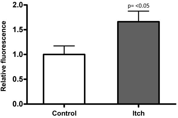
Figure 4.
Itch regulation TGFβ signaling, as determined by PAI-1/luciferase activity in mink lung epithelial cells (seeMethods). Cells were either transiently transfected with pCDNA3.1 empty vector (control) or mouse Itch cDNA in pCDNA3.1 under the control of a CMV promoter. Both groups were treated with TGFβ (200 ng/ml, 8 hr, 37°C). The readout (relative fluorescence), which is depicted on the Y-axis is calculated from the ratio of fluorescence units in cells transfected with Itch to fluorescence units in control cells (n = 3). Results are expressed as mean ± SD. A two-tailed t-test yielded a p-value of < 0.05 between groups.

