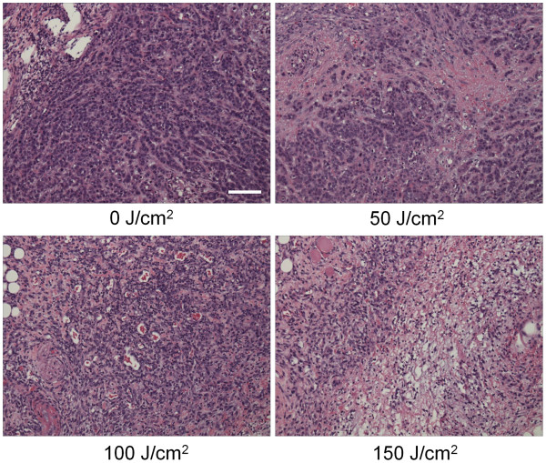
Figure 4 .
Histological findings immediately after PIT with various NIR exposures. Histological specimens of MDA-MB-468luc tumors, which were treated with PIT at 0, 50, 100, and 150 J/cm2, are shown. All specimens are stained with Hematoxylin and Eosin. A few scattered clusters of damaged tumor cells are seen within a background of diffuse cellular necrosis and micro-hemorrhage immediately after PIT with 50 J/cm2. Necrotic damage was more intense when higher intensity NIR light was administered. Scale indicates 50μm.

