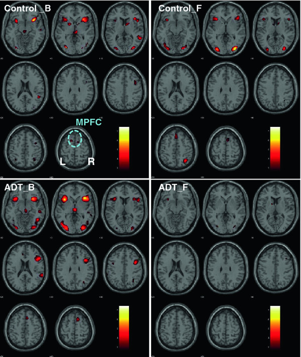
Figure 1.
Regional brainactivations duringcognitive controlwhile performingthe stopsignal task. ADT = patients who received 6 months of androgen deprivation therapy; Controls = patients who did not receive any hormonal therapy; B = baseline; F = at 6- month follow-up. The significance of activation, as reflected by a map of T values (color bar), is shown here on a structural brain image in axial sections, from z = -10 to z = +60, with adjacent sections 10 mm apart. Neurological orientation: Right (R) = right. Note that ADT patients showed diminished activations in a number of brain regions at 6 month follow-up, including the medial prefrontal cortex (MPFC), an area critical for cognitive control.

