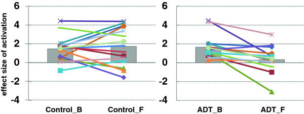
Figure 2.
The effectsize ofmedial prefrontalcortical (MPFC)activations. Control patients are shown on the left panel; ADT patients are shown on the right panel; B = baseline; F = at 6-month follow-up. In each panel, each symbol and line represents the data of an individual patient. The gray bars indicate the mean value of the effect sizes of MPFC activations. Individuals varied in the change of MPFC activations from baseline to follow-up, but, on average, the ADT group showed significant decrease in MPFC activations after 6 months of ADT, as compared to the control group.

