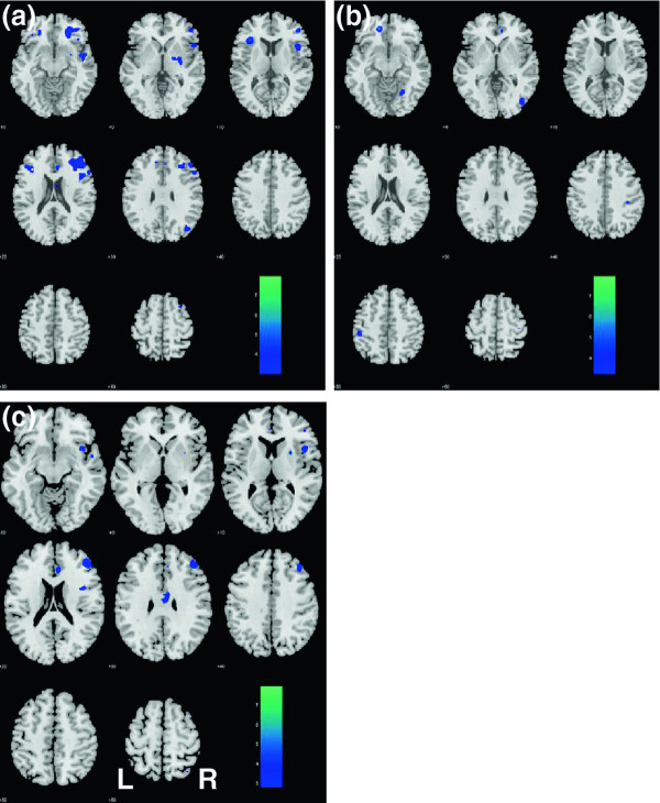
Figure 3.
Changes inresting statefunctional connectivitywith themedial prefrontalcortex asresult ofADT. After 6 months of ADT, patients showed decreased connectivity with the dorsolateral prefrontal cortex (DLPFC), rostral anterior cingulate cortex, and the insula, all on the right side, as compared to control patients. (a) ADT_B > ADT_F; (b) Control_B > Control_F; (c) (Control_F > Control_B) – (ADT_F > ADT_B). B: baseline; F: follow-up. The significance of difference, as reflected by a map of T values (color bar), is shown here on a structural brain image in axial sections, from z = -10 to z = +60, with adjacent sections 10 mm apart. Neurological orientation: Right (R) = right.

