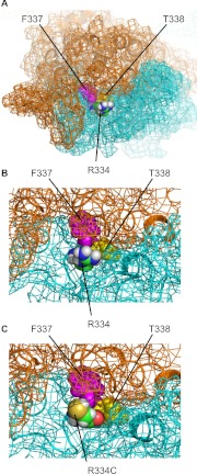
Fig. 9.
Homology model of the CFTR based on MsbA (Mornon et al., 2009). A, top view from the extracellular side. Arg334, Phe337, and Thr338 are shown as van der Waals representations. The carbon atoms of Thr334 are colored green, and Phe337 and Thr338 are colored pink and yellow, respectively. B, close-up view of the structure shown in A, around Thr334. The CFTR molecule is shown as a cartoon representation, with its molecular surface indicated by mesh. Transmembrane domain 1 is shown in cyan and transmembrane domain 2 in orange. C, view with Thr334, shown in B, mutated to cysteine with Maestro 9.1 (Schrödinger).

