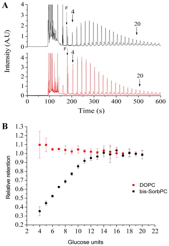
Figure 3.
Size-based retention of dextrans in PPNs. (A) Electropherograms of APTS-labeled dextran retained within PPNs (top) and DOPC vesicles (bottom). Peaks corresponding to dextrans comprised of 4 and 20 glucose units are denoted by arrows. (B) Relative retention as a function of size for dextrans retained in DOPC vesicles (squares) and PPNs (circles).

