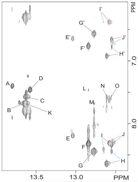
Figure 4.
Contour plot of an expanded region of an 800 MHz NOESY (220 ms mixing time) spectrum recorded in 10% D2O buffer at 5 °C, showing interactions between the imino and base/amino proton regions. Red and blue colors differentiate the Z and E isomeric duplexes, respectively. Labeled peaks are assigned as follows: A, T19(N3H) A4(H2); B, T9(N3H) A14(H2); C, T15(N3H) A8(H2); D, T3(N3H) A20(H2); E/E′, G12(N1H) C11(N4H)hb/nhb; F/F’, G2(N1H) C21(N4H)hb/nhb; G/G’, G10(N1H) 13(N4H)hb/nhb; H/H’, G16(N1H) C7(N4H)hb/nhb; I/I’, F(N3H) C17(N4H)hb/nhb; J/J’, G18(N1H) C5(N4H)hb/nhb; K, T15(N3H) A14(H2); L, G2(N1H) A20(H2); M, G10(N1H) A14(H2); N, G16(N1H) A8(H2); O, F(N3H) F(N2H).

