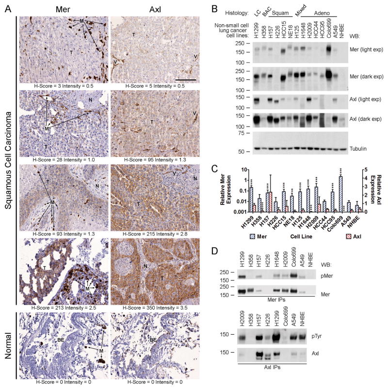
Figure 1. Mer and Axl receptor tyrosine kinases are expressed in human non-small cell lung cancer tumors and cell lines.
(A) Immunohistochemical staining of Mer and Axl in sections of normal human lung and squamous cell carcinoma of human lung. Micrographs shown represent the range of staining observed in tumor cells (T). Adjacent normal tissue (N) was always negative for Mer and Axl as was bronchial epithelium (BE). Macrophages (M, arrows) were strongly positive for Mer and blood vessels (V) were strongly positive for Axl. The scale bar in the upper right panel is applicable to all micrographs and represents 100 μm. (B) Whole-cell lysates of the indicated cell lines were subjected to western blot (WB) analysis using the indicated antibodies. Two exposures (exp) are shown for Mer and Axl. In 13/13 NSCLC lines tested, Mer and/or Axl are overexpressed relative to levels found in normal human bronchial epithelial (NHBE) cells. Variation in the molecular weight of each receptor is likely due to cell line-specific patterns of glycosylation as the extracellular domains of Axl and Mer contain 6 and 13 glycosylation sites, respectively. Tubulin was used as a loading control. Numbers on the left indicate the location of molecular weight (kDa) markers. Blots representative of at least three independent experiments are shown. (C) Real-time RT-PCR analysis of Mer and Axl mRNA transcript expression in the same collection of cell lines shown in panel B. Two-way ANOVA and Bonferroni post-tests were used to compare the mean ΔCt values derived from at least three independent measurements. The mean + SD of antilogged ΔCt values (2−Δ Ct) are shown. The asterisks indicate significant differences versus NHBE (*** P < 0.001, ** P < 0.01, * P < 0.05). (D) Mer and Axl were immunoprecipitated from whole-cell lysates of steady-state cultures of a subset of the NSCLC cell lines shown in A and B. Phosphorylated and total levels of Mer and Axl proteins were assessed by western blot analysis using anti-phospho-Mer (pMer, Y749/Y753/Y754, PhosphoSolutions) and anti-Mer antibodies or anti-phosphotyrosine (pTyr, Millipore, 05–1050) and anti-Axl antibodies, respectively. Blots representative of at least three independent experiments are shown.

