
Figure 1.
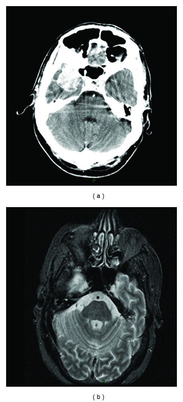
CT scan (a) and MRI (b) revealed a 4.5 × 4.5 × 3-cm mass that enhanced with contrast, in the right greater wing of the sphenoid bone which was invading the anterior pole of the temporal lobe and the sphenoid sinus.
Official websites use .gov
A
.gov website belongs to an official
government organization in the United States.
Secure .gov websites use HTTPS
A lock (
) or https:// means you've safely
connected to the .gov website. Share sensitive
information only on official, secure websites.
CT scan (a) and MRI (b) revealed a 4.5 × 4.5 × 3-cm mass that enhanced with contrast, in the right greater wing of the sphenoid bone which was invading the anterior pole of the temporal lobe and the sphenoid sinus.