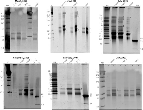
Fig 1.
Pulsed-field gels of virioplankton concentrates. Gels are labeled according to the month and year of sample collection. Lanes are labeled by station designation in the Chesapeake Bay (CB) and Delaware Bay (DB). Samples from the June 2006 and July 2007 cruises were loaded at two different concentrations of virus particles: 109 (A) and ∼1010 (B). DNA was purified from bands marked with numbers. Marker lanes (in kilobases) are as follows: M1, concatemers of phage λ genome mixed with HindIII digest of λ genomic DNA; M2, concatemers of phage λ genome; M3, λ DNA digested with HindIII; M4, midrange PFG marker.

