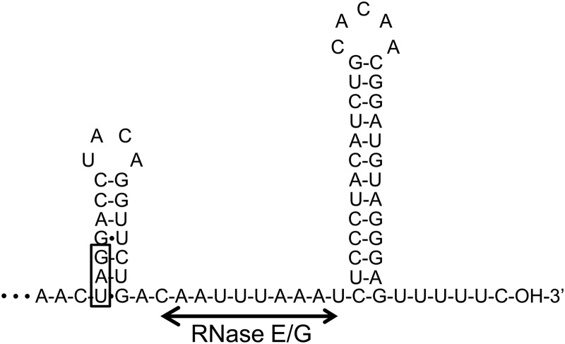
Fig 5.
Predicted secondary structure of the aceA 3′-UTR. RNA fold software (mfold [http://mfold.rna.albany.edu/?q=mfold]) was used to predict the secondary structure of the aceA 3′-UTR. The UAG stop codon is boxed. The horizontal line represents the possible cleavage site of RNase E/G (see the text for details).

