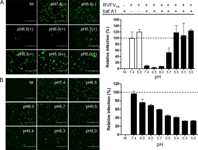
Fig 3.
Low-pH-activated penetration of cell-bound RVFVns and inactivation of unbound RVFVns particles. (A) RVFVns was allowed to bind in the cold to a confluent monolayer of BHK-21 cells for 2 h before the cell-bound virus was exposed for 3 min at 37°C to the indicated pH. Infection was continued in culture medium containing bafilomycin A1 (baf A1; 20 nM) to inhibit infection via the endocytic route. Infection (GFP-positive cells) was analyzed 20 h after warming by fluorescence microscopy and was quantified by FACS. Untreated cells (pH 7.4) yielded ∼40% infection. The results shown are representative of two individual experiments performed in triplicate. (B) RVFVns particles were incubated at the indicated pH for 3 min at 37°C. After neutralization of the medium, infectivity of the virus was assayed on BHK-21 cells. Infection (GFP-positive cells) was analyzed 20 h.p.i. by fluorescence microscopy (left panel; size bars represent 400 µM) and quantified by FACS (right panel). Virus incubated at neutral pH resulted in ∼30% GFP-positive cells. Graphical data shown are representative of four individual experiments performed in duplicate. The controls in the experiments whose results are shown in panels A and B were cells that were not infected (NI).

