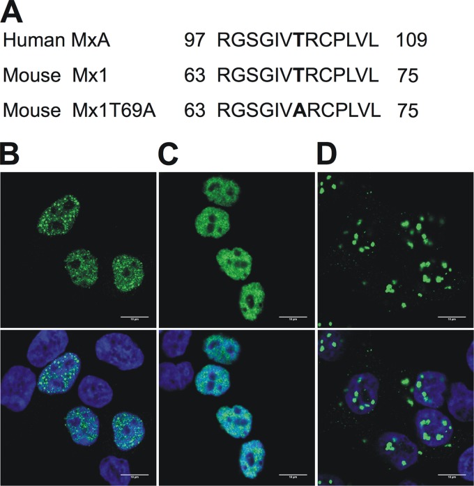
Fig 4.
GTPase-deficient Mx1 mutants. (A) Alignment of the amino acid sequence of the human MxAT103A mutant, wild-type mouse Mx1, and the mouse Mx1T69A mutant. The mutation is localized between the first and second GTP-binding consensus motifs. Numbers denote the position in the primary amino acid sequence. (B to D) Subcellular localization of Mx1 (B), Mx1K49A (C), and Mx1T69A (D). HEK293T cells were transfected with pCAXL-Mx1WT, -Mx1K49A, or -Mx1T69A (50 ng). After 24 h, cells were fixed and stained with Hoechst stain (DNA, blue, not shown) and anti-Mx1 antibody (green, top). An overlay was made of the two images (bottom). Scale bar, 10 μm.

