
Fig 3.
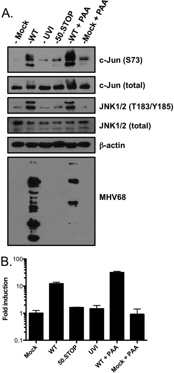
JNK/c-Jun phosphorylation and AP-1 reporter activity are amplified as infection progresses. (A) Serum-starved 3T3 fibroblasts were either mock infected or infected at an MOI of 5 PFU/cell with WT MHV68, UVI MHV68, ORF50-null MHV68 (50.STOP), mock infected, or infected with WT MHV68 in the presence of PAA (200 μg/ml). Cells were harvested 18 h postinfection, and proteins were resolved by SDS-PAGE. Resolved proteins were detected by immunoblot analyses using antibodies that recognize the indicated proteins. (B) 3T3 fibroblasts were transiently transfected with an AP-1 reporter plasmid to evaluate JNK/c-Jun transcriptional activity. At 24 h posttransfection, cells were replated and infected as described for panel A. Cells were harvested 18 h postinfection, and luciferase activity was quantified. Results are means of triplicate samples. Error bars represent standard deviations.