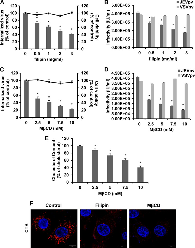
Fig 5.
Filipin and MβCD inhibit JEV entry into B104 cells. (A and C) B104 cells were pretreated with filipin (A) or MβCD (C) and infected with JEV. After 1 h of internalization, extracellular virus was inactivated with proteinase K and the cell pellets were plated onto B104 cells to determine internalized virus by an infectious center assay. Bar graphs represent the percentage of internalized virus with respect to a control without drug treatment. Cell viability upon drug treatments was unaffected, as represented by the line graphs. Results are presented as the means ± SD of three independent experiments. (B and D) B104 cells preincubated with filipin (B) or MβCD (D) were inoculated with JEVpv or VSVpv. GFP-positive cells were determined by flow cytometry. Results are expressed as infectious units (IU)/milliliter and are presented as the means ± SD of three independent experiments. (E) Intracellular cholesterol levels were quantitated in mock-treated or MβCD-treated B104 cells and compared to those in mock-treated B104 cells. Results are expressed as the means ± SD of three independent experiments. (F) B104 cells were left untreated (control) or were treated with 1 mg/ml filipin or 10 mM MβCD for 1 h at 37°C and then incubated with 10 μg/ml AF 555-labeled cholera toxin B (CTB) for 30 min at 37°C. Nuclei were stained with DAPI. Bars, 10 μm. *, P < 0.05.

