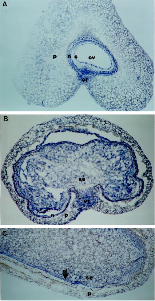
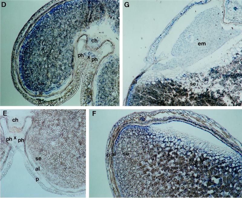
Figure 5.
(Figure continues on facing page.)
Immunological localization of PEPC in developing wheat grains. Immunolocalizations were carried out as described in Methods. Sections of grains (10 μm thick) 5 DPA (A), 10 DPA (B and C), 16 DPA (D and F), and 22 DPA (G) were probed with polyclonal PEPC IgGs (1 μg of protein per slide). A control section from 16-DPA grains was probed with preimmune serum (E). al, Aleurone layer; ch, chalaza; cv, central vacuole; em, embryo, n, nucellus; p, pericarp; ph, phloem; s, syncytium; se, starchy endosperm; vt, vascular tissue; and x, xylem. Magnification ×50.


