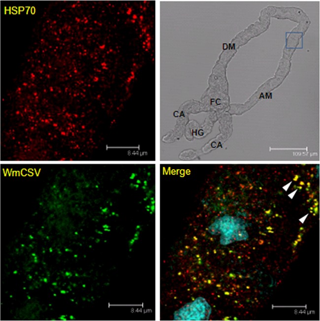
Fig 7.
Colocalization of HSP70 and WmCSV in midguts of viruliferous whiteflies. HSP70 immunolocalization using specific first antibody and secondary antibody conjugated to Alexa Fluor (635 nm) (red) and WmCSV immunolocalization using specific first antibody and secondary antibody conjugated to Alexa Fluor (488 nm) (green). Note the yellow areas indicating colocalization of WmCSV and HSP70 in the ascending midgut (AM). Blue indicates DAPI staining of the nuclei. DM, descending midgut; CA, cecae; HG, hindgut; FC, filter chamber.

