Fig 1.
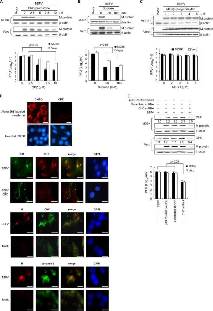
BEFV infection impaired by inhibition of clathrin-coated pit formation. (A and B) MDBK and Vero cells were pretreated with various concentrations of CPZ (A) and hypertonic sucrose (B) for 1 h, followed by infection with BEFV at an MOI of 2. The level of M protein and the progeny virus titer of BEFV were examined by Western blotting and by plaque assay, respectively. (C) MDBK and Vero cells were pretreated with various concentrations of MβCD for 1 h, followed by infection with BEFV at an MOI of 2. The cell lysates and supernatants of BEFV-infected cells were collected at 24 hpi for Western blotting and viral titration, respectively. The level of M protein was detected by Western blotting. The progeny virus titer of BEFV was determined by plaque assay. The results are from three triplicate experiments; error bars indicate the means ± standard deviations. The β-actin was used as an internal control for normalization. (D) Fluorescence of Alexa 568-labeled transferrin (Tfn) is shown, along with a Hoechst 33258 counterstain for cell nuclei. To study whether CPZ affects virus internalization, MDBK cells were starved for 2 h in serum-free medium and then were pretreated with CPZ (5 μM) for 1 h. MDBK cells were then infected with DiO-labeled BEFV. DiO-labeled viral particles were observed with a fluorescence microscope. Colocalization of the M protein of BEFV with clathrin or dynamin 2 was also observed by fluorescence microscopy. Immunostaining of clathrin, dynamin 2, and BEFV M was performed using respective antibodies. Mock-infected cells were used as negative controls. Cell nuclei were counterstained with 4′,6-diamidino-2-phenylindole (DAPI). Bars, 25 μm. (E) BEFV infection is inhibited in cells transfected with shRNAs specific to clathrin heavy chain. MDBK and Vero cells were transfected with CHC shRNA and mock control vectors (scrambled pGFP-V-RS and pGFP-V-RS), respectively. MDBK and Vero cells were infected with BEFV at an MOI of 2. The cell lysates and supernatants of BEFV-infected cells were collected at 24 hpi for Western blotting and viral titration, respectively. The β-actin was included as an internal control for normalization. Numbers below each lane are percentages of the control level of a specific protein in mock-treated cells. The results are from three triplicate experiments; error bars indicate the means ± standard deviations.

