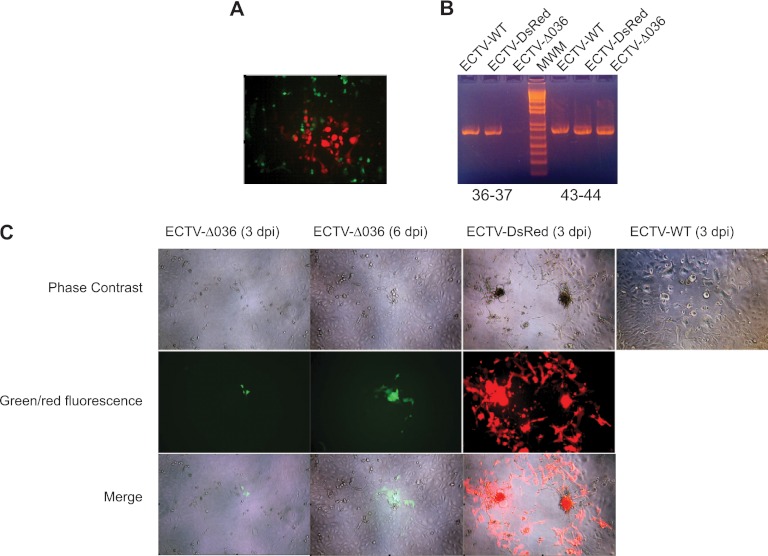
Fig 2.
Identification of ECTV-Δ036 and ECTV-DsRed. (A) Microphotograph showing a red ECTV-DsRed plaque within ECTV-Δ036 (green)-infected cells 2 days after infection of B-SC-1 cells with a lysate of B-SC-1 cells infected with ECTV-Δ036 and transfected with pBSSK-RevΔ036-DsRed. (B) PCR analysis of lysates of the indicated viruses using primers to amplify the junction of EVM036 and EVM037 or the junction of EVM43 and EVM44. (C) Monolayer of B-SC-1 cells infected with the indicated viruses and visualized by microscopy on the indicated day postinfection.

