
Figure 6.
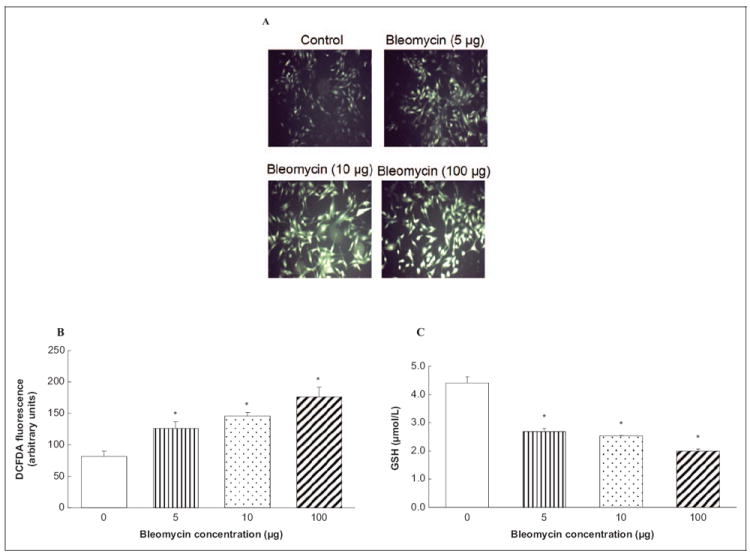
Bleomycin induces ROS generation and depletes GSH in ECs. Bovine lung microvascular ECs ([BLMVECs 5 × 10⁵ cells/35-mm dish) were preloaded with 10 μmol/L DCFDA for 30 minutes in complete MEM to determine ROS generation. Following the DCFDA loading, cells were subjected to treatment with MEM alone or MEM containing bleomycin (5, 10, and 100 μg) for 1 hour (A) and 0.5 hour (B). At the end of the incubation period, the DCFDA fluorescence (as an index of ROS formation) was determined as described under Materials and Methods section. Each micrograph is a representative picture obtained from 3 independent experiments conducted under identical conditions. Bovine lung microvascular ECs (1 × 10⁵ cells/96 well plates) were subjected to treatment with MEM alone or MEM containing bleomycin (5, 10, and 100 μg) for 12 hours to determine intracellular GSH levels (C). At the end of the incubation period, the intracellular soluble thiol (GSH) concentrations were determined. Data represent mean ± SD calculated from 3 independent experiments. *Significantly different at P < .05 as compared to cells treated with MEM alone. DCFDA indicates 2′-7′-dichlorofluorescin diacetate; MEM, minimal essential medium; ROS, reactive oxygen species; GSH, glutathione; ECs, endothelial cells.