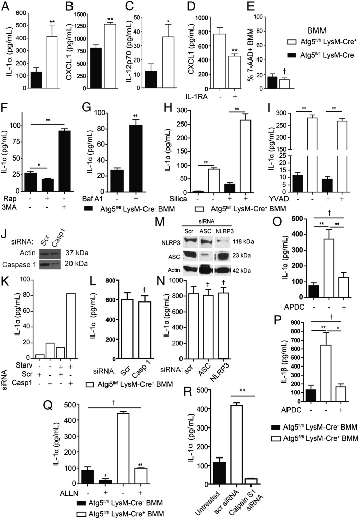
Fig. 5.
Excess cytokine secretion is a cell-autonomous property of autophagy-deficient macrophages, and IL-1α hypersecretion by Atg5fl/fl LysM-Cre+ macrophages depends on reactive oxygen intermediates and calpain. (A–C) In vitro cytokine [IL-1α (A), CXCL1 (B), and IL-12p70 (C)] release (ELISA) from LPS- and IFN-γ–stimulated Atg5fl/fl LysM-Cre− and -Cre+ BMM. (D) CXCL1 released (ELISA) from LPS- and IFN-γ–stimulated Atg5fl/fl LysM-Cre+ BMM in the absence of presence of IL-1RA (0.5 µg/mL). (E) Fraction (flow cytometry) of 7-AAD+ BMM after LPS and IFN-γ stimulation in vitro. (F and G) IL-1α (ELISA) released from LPS- and IFN-γ−stimulated Atg5fl/fl LysM-Cre− BMM in the presence of 50 μg/mL rapamycin (Rap) or 10 mM 3-MA (F) or 100 nM Baf A1 (G) after 12 h of treatment. (H) IL-1α secretion during inflammasome activation. Atg5fl/fl LysM-Cre− and -Cre+ BMM were pretreated overnight with LPS (100 ng/mL) and then were stimulated for 1 h in the absence or presence of the inflammasome agonist silica (250 μg/mL) in EBSS. (I) IL-1α secretion in the presence of caspase 1 inhibitor YVAD. Atg5fl/fl LysM-Cre− and -Cre+ BMM were pretreated overnight with LPS (100 ng/mL) and then were stimulated for 1 h in the absence or presence of YVAD (50 μM) during inflammasome activation with silica as in H. (J–L) Effects of caspase 1 siRNA knockdown (immunoblot, J) on IL-1α release (graphs, K and L) from Atg5fl/fl LysM-Cre+ BMM. (K) IL-1α release was measured (ELISA) from LPS-stimulated and siRNA-treated Atg5fl/fl LysM-Cre+ BMM in full medium or EBSS (Starv) (K) or in full medium only (L). Casp 1, caspase 1 siRNA; Scr, scrambled siRNA (control). (M and N) Effects of NLRP3 and ASC siRNA knockdown (immunoblot, M) on IL-1α release (N). IL-1α release was measured (ELISA) from LPS- and IFN-γ–stimulated Atg5fl/fl LysM-Cre+ BMM knocked down with siRNA for inflammasome components ASC and NLRP3. (O and P) ROS inhibition and IL-1 secretion. IL-1α (O) and IL-1β (P) released (ELISA) from LPS- and IFN-γ–stimulated Atg5fl/fl LysM-Cre− and -Cre+ BMM in the absence or presence of the ROS antagonist APDC (50 μM) after 12 h of incubation. (Q) Calpain and IL-1α hypersecretion phenotype. IL-1α (ELISA) released from LPS- and IFN-γ–stimulated Atg5fl/fl LysM-Cre− and -Cre+ BMM in the absence or presence of the calpain inhibitor ALLN (100 μM) after 12 h of stimulation. (R) IL-1α release from LPS- and IFN-γ–stimulated Atg5fl/fl LysM-Cre− BMM and Atg5fl/fl LysM-Cre+ BMM knocked down with siRNA for Calpain S1. Data are shown as mean ± SE; *P < 0.05, **P < 0.01, †P > 0.05 (t test; n ≥ 3).

