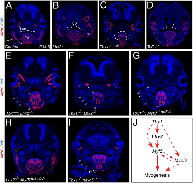
Fig. 4.
Epistatic genetic relationships regulating pharyngeal muscle development. Transverse craniofacial sections of E14.5 mouse embryos stained with MyHC for single- (B–D) and double- (E–J) mutants for the indicated genotypes (n ≥ 4). Dotted line outlines the cleft palate seen in all three single mutants (B–D). (J) A model summarizing the genetic interactions described above. Dotted arrows indicate parallel regulatory interactions affecting head myogenesis, empty arrowheads indicate novel interactions. Skeletal muscle groups are marked in white arrowheads, and their absence in black arrowheads. first/second, first/second pharyngeal arch-derived muscles; tng, tongue.

