Fig. 1.
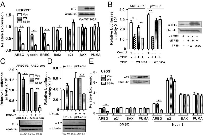
The integrity of TFIIB ser65 is not critical for the expression of p53-target genes. (A) qPCR analysis of AREG, γ-actin, EREG, Bcl2, p21, BAX, and PUMA gene expression was carried out after 48 h of transfection with pCDNA3 vector, T7-tagged WT TFIIB and TFIIB S65A in HEK293T cells. Western analysis with anti-T7 antibody confirmed equivalent expression of WT TFIIB and TFIIB S65A. (B) Luciferase activity of FL promoter constructs of AREG and p21 was measured in cells transfected with pSUPER shTFIIB along with WT or S65A TFIIB. Transfection with empty pSUPER vector and shTFIIB are controls. Error bars denote SD of three independent experiments. Western blotting analysis was performed with anti-TFIIB and anti-β tubulin antibodies. (C) Luciferase assay was performed after pCDNA3 vector, T7-tagged WT TFIIB and TFIIB S65A transfection with FL promoter or core promoter constructs of AREG and (D) p21 promoter constructs. Error bars denote SD of three independent experiments. Western blotting analysis was as above. (E) qPCR analysis of AREG, p21, BAX, and PUMA gene expression was carried out in U2OS cells transfected with pCDNA3 vector, T7-tagged WT TFIIB and TFIIB S65A for 40 h followed by 8 h of treatment with 10 μM Nutlin3 or DMSO. Western analysis was as above.

