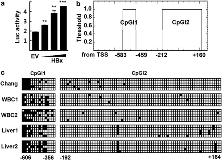
Figure 1.
Methylation analysis of the MTA1 promoter. (a) HepG2 cells were transfected with the human MTA1 promoter-Luc reporter and increasing amounts of p3XFLAG7.1-HBx or empty vector (EV). Experimental values are expressed as the mean±s.d. (n=3). **P<0.01 and ***P<0.001 vs EV transfected. (b) Putative CpG islands are located at bases −583 to −459 (CpGI1) and −212 to +160 (CpGI2) in the human MTA1 promoter. (c) Sequencing analysis of the putative MTA1 promoter CpG islands after bisulfite modification. Five clones from Chang liver cells (Chang), human white blood cells (WBC1 and WBC2), and normal liver tissues (Liver1 and Liver2) were analyzed for each PCR fragments. Filled squares, methylated; and open squares, unmethylated.

