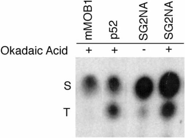
Fig. 4. mMOB1 is phosphorylated only on serine residues, whereas SG2NA and p52 are phosphorylated on both serine and threonine.
NIH3T3 cells were labeled in vivo with [32P]orthophosphate and either treated (1) with 1 mm okadaic acid or left untreated (–). SG2NA complexes were immunoprecipitated and analyzed by SDS-PAGE, and phosphoamino acid analysis was performed as described (38). In the absence of okadaic acid, mMOB1 and p52 cannot be detected with 32P and, thus, are not shown. Phosphoserine residues (S) and phosphothreonine residues (T) are indicated on the left of the figure. No phosphotyrosine was detected in any of these proteins (not shown).

