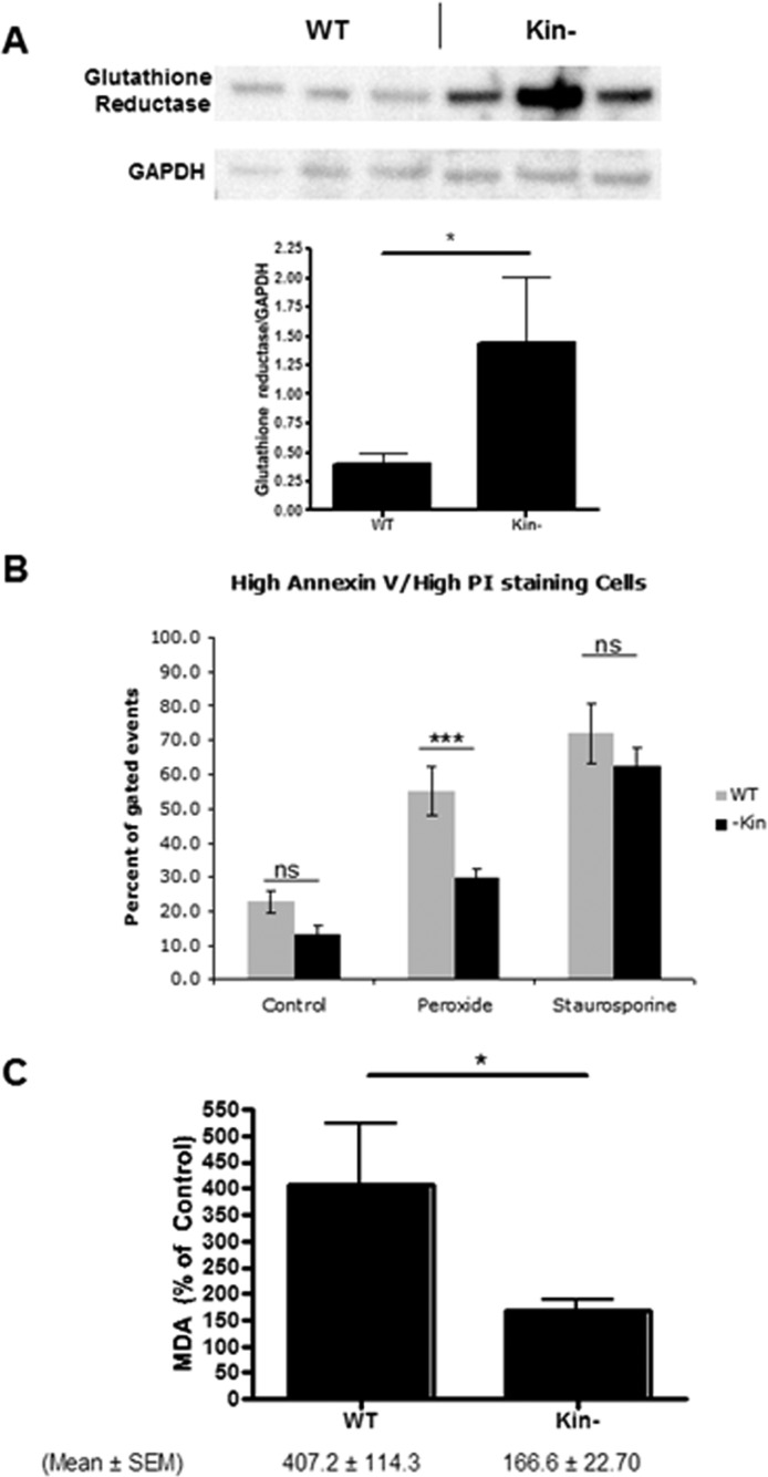
Figure 2.
Enhanced glutathione reductase expression and reduced sensitivity to cell death and lipid peroxidation in response to H2O2 of kin- vs WT S49 cells. (A) Immunoblotting confirms data from the proteomics analysis that kin- cell lysates have more glutathione reductase than do WT S49 cell lysates (both 25 μg of protein/well). The bottom panel shows quantitation of protein expression by densitometry (mean ± SEM; n = 3). *p < 0.05. (B) Incubation of WT and kin- cells for 16 h at 37 °C with 150 μM H2O2 produced greater cell death (representing both necrosis and apoptosis) in WT cells than in kin- cells, but this difference was not observed in cells incubated with 1 μM staurosporine. kin- cells also experienced less cell death under control conditions (incubation with H2O for 16 h). (C) A 16 h incubation at 37 °C with 150 μM H2O2 produced more lipid peroxidation, assessed as MDA, in WT cells than in kin- cells. The data shown in panels B and C are means ± SEM (n = 7). *p < 0.05; ***p < 0.001.

