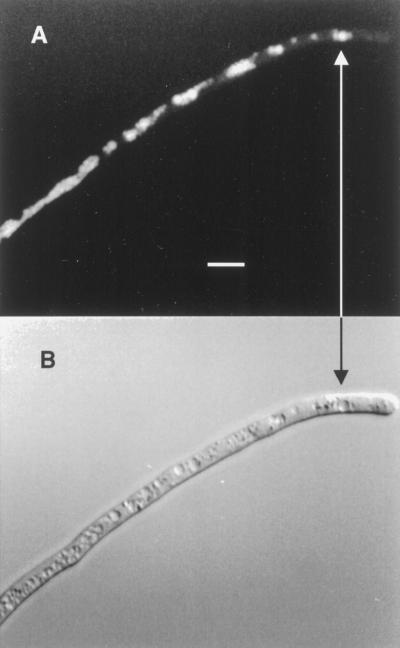
Figure 2.
Senescing G. margarita germ tube 3 h after BCECF-AM loading into a lethal DMSO concentration. A, BCECF fluorescence microscopy (450 nm excitation). B, Same hypha obtained by differential interference contrast microscopy. The fluorescence localizes in cytoplasmic areas not yet necrosed (arrow). Bar = 10 μm.

