Figure 4.
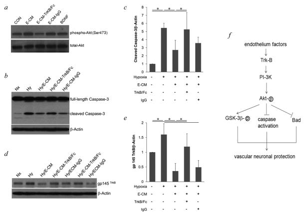
a, E-CM depleted by TrkB/Fc did not induce the phosphorylation of Akt in neurons after 30 min incubation. b, incubation with E-CM blocked cleavage of Caspase-3 in neurons after hypoxia, but incubation with E-CM depleted by TrkB/Fc had no effect on increased cleavage of Caspase-3 in neurons after hypoxia. c, optical densitometry of cleaved Caspase-3, normalized to β–Actin. d, hypoxia increased TrkB expression in neurons, which was blocked by E-CM. Depleted E-CM had no blocking effect on TrkB expression. e, optical densitometry of gp 145 TrkB, normalized to β–Actin. * P<0.05 between two groups. f, proposed mechanisms for neuroprotection by cerebral endothelial cells. Soluble factors from cerebral endothelial cells, such as growth factor BDNF, activate PI-3K/Akt survival signaling in neurons through receptors, leading to the activation of GSK-3β, the inhibition of caspases activation and Bad expression, and finally protect the neurons from damage.

