Fig. 4.
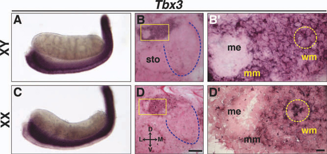
Expression of Tbx3 in Wolffian and Müllerian ducts at E13.5. Whole-mount ISH and section ISH on transverse sections of XY (A, B, B′) and XX (C, D, D′) embryos. In XY and XX embryos, Tbx3 is expressed in the Wolffian duct epithelium (dashed yellow circles) and mesenchyme around Wolffian and Müllerian ducts, but not in testes (A, B) or ovaries (C, D). Boxed areas in B and D are shown at higher magnification in B′ and D′. Dashed blue lines outline gonads in transverse sections. D, dorsal; L, lateral; M, medial; V, ventral; me, Müllerian duct epithelium; mm, Müllerian duct mesenchyme; sto, stomach; wm; Wolffian duct mesenchyme. Scale bar in D = 100 µm (B, D); scale bar in D′ = 10 µm (B′, D′).

